
Figure 24 (A, B).
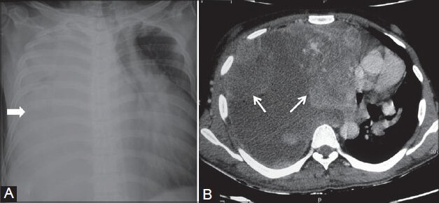
Spindle cell sarcoma of pleura: (A) Chest radiograph showing complete opacification of right hemithorax (arrowhead) with mediastinal shift to the left; (B) axial contrast-enhanced CT scan showing heterogeneously enhancing nodular pleural-based lesions with pleural effusion displacing the heart to the left