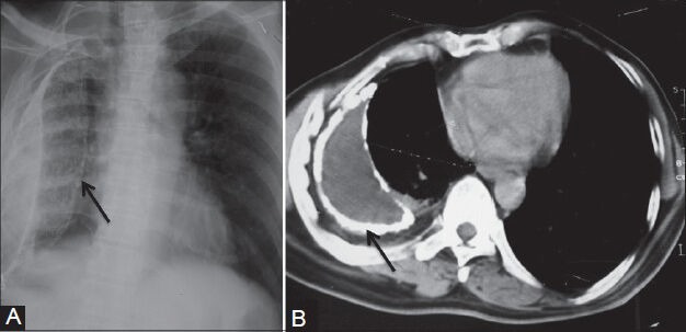
Figure 4 (A, B).

Calcified empyema: (A) Chest radiograph showing volume loss right hemithorax with veil-like calcified (arrow) pleural opacity; (B) axial contrast-enhanced CT scan showing evidence of calcified chronic empyema (arrow) with proliferation of extrapleural fat and crowding of ribs suggestive of volume loss in right hemithorax
