
Figure 6 (A, B).
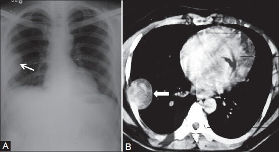
Benign solitary fibrous tumor: (A) Chest radiograph showing pleural-based opacity (arrow) in right hemithorax with peripheral obtuse margins; (B) axial contrast-enhanced CT scan showing heterogeneously enhancing pleural-based mass (arrowhead) proved to be benign fibrous pleural tumor