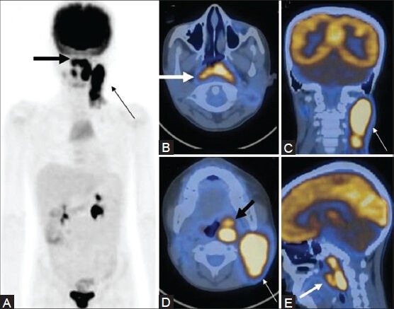
Figure 16 (A-E).

A case of NHL showing nasopharyngeal involvement extending inferiorly into the oropharynx (thick arrow), with bulky cervical lymphadenopathy (thin arrow) on the MIP (A) and transaxial , coronal, and sagittal PET/CT fusion images (B-E)
