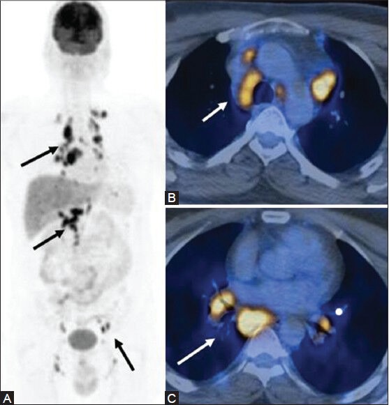
Figure 22 (A-C).

A case of disseminated Koch's masquerading as lymphoma: MIP image (A) Multiple sites of lymph node involvement (arrows). Transaxial PET/CT fusion images (B, C) sites of increased FDG uptake in the enlarged mediastinal and hilar lymph nodes
