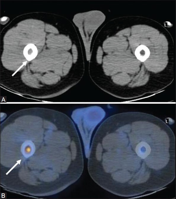
Figure 3 (A, B).

Transaxial CT image at the level of upper thigh (A) In a case of NHL (DLBCL) appears unremarkable. PET/CT fusion image at the corresponding site (B) intense FDG uptake in the bone marrow of proximal right femoral shaft (arrow) suggestive of marrow involvement
