Figure 12.

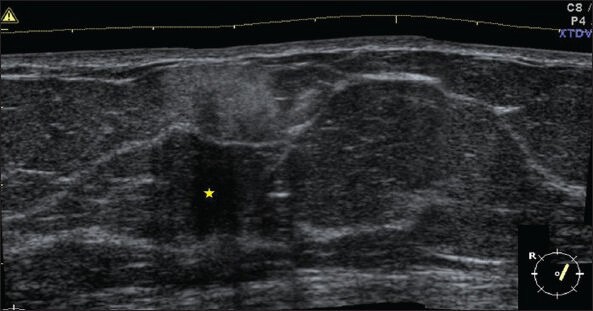
USG image of cytologically proven fat necrosis shows superficially located ill-defined hyperreflective lesion with marked posterior acoustic shadowing (*)

USG image of cytologically proven fat necrosis shows superficially located ill-defined hyperreflective lesion with marked posterior acoustic shadowing (*)