Figure 13.

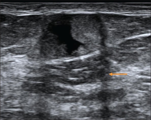
Follow-up USG image of the lesion shown in Figure 8 at 3 months, which shows anechoic component within the lesion. Edge shadowing seen here (arrow) is neither a benign nor a malignant feature

Follow-up USG image of the lesion shown in Figure 8 at 3 months, which shows anechoic component within the lesion. Edge shadowing seen here (arrow) is neither a benign nor a malignant feature