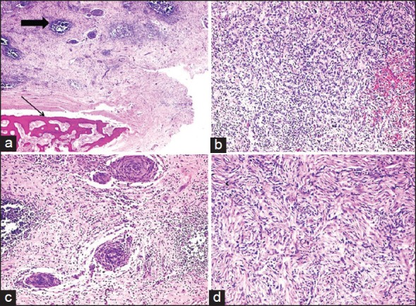
Figure 1.

Low power view of inflammatory myofibroblastic tumor (IMFT) of kidney with heterotopic bone formation (thin arrow). Even at this power the lymphoid aggregates are prominent (thick arrow), (a) (H and E, ×40). The presence of spindle cells, inflammatory cells with extravasated red blood cells, (b) (H and E, ×100), perivascular whorls of spindle cells, (c) (H and E, ×100) and focal storiform pattern, (d) (H and E, ×200) was demonstrable in various areas of IMFT
