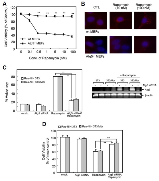
Fig. 2. Effect of rapamycin on viability and autophagy of WT and Atg5-/- MEFs. (A) Autophagy-proficient (WT MEFs) and -deficient (Atg5-/- MEFs) cells were treated with increasing concentrations of rapamycin ranging from 0.1 to 100 nM for 72 h. Cell viability was evaluated with WST-1 reagent. The viability of cells treated with vehicle alone was regarded as 100%. Values represent the mean ± SD of quadruplicate determinants from one of three representative experiments. **P < 0.01 as determined by the Dunnett’s T-test as compared to drug-sensitive cells. (B) Autophagy induction was measured by immunofluorescent staining using anti-LC3 (Red). Blue = DAPI staining. (C) Ras-NIH 3T3/Mdr cells were transiently transfected with Atg5 siRNA or scrambled control siRNA for 24 h, together with GFP-LC3. Cells were subsequently washed and then treated with or without rapamycin (10 nM) for 24 h. Autophagy was quantified by the GFP-LC3 puncta. Right inset, RT-PCR showing knock-down of Atg5 mRNA expression by Atg5 siRNA. (D) Atg5 knockdown cells were incubated in the presence or absence of rapamycin in 96-well plates for 3 days. Cell growth inhibition was eva-luated with WST-1 reagent. In (C) and (D), the value of mock-transfected cells was regarded as 100%. Values represent the mean ± SD of quadruplicate determinants from one of three representative experiments. **P < 0.01 as determined by Dunnett’s t-test.