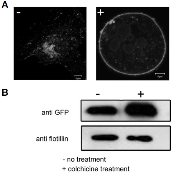
Fig. 4. Translocation of mMARVELD1 to the plasma membrane after treatment with colchicine. (A) NIH3T3 cells expressing mMARVELD1- EGFP were treated with 1 mg/ml colchicine (right panel). 12 h after treatment, the cells were observed by confocal microscopy. (B) The plasma membrane fraction from colchicine-treated or control NIH3T3 cells expressing mMARVELD1-EGFP was extracted, and the amount of mMARVELD1-EGFP in the plasma membrane fraction was detected by Western blot using anti-GFP antibody. The membrane protein flotillin was used as plasma membrane marker.

