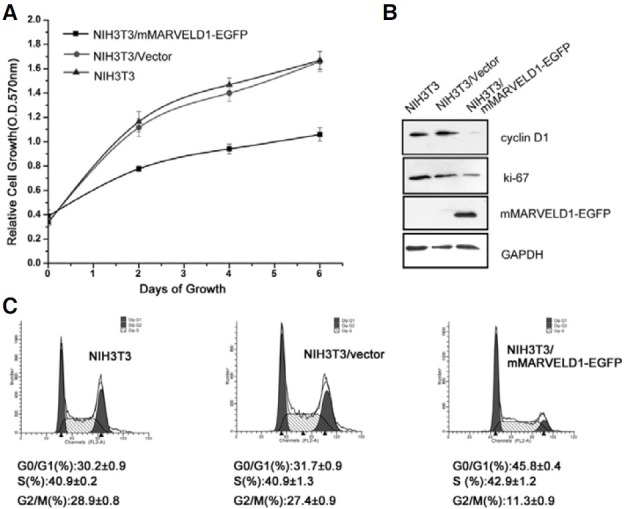
Fig. 5. Overexpression of mMARVELD1 inhibits NIH3T3 cell proliferation. (A) NIH3T3 cells expressing mMARVELD1-EGFP or control vector and untransfected cells were seeded into 96-well plates. Cell proliferation was measured by MTT assay at days 0, 2, 4, and 6. (B) Cell lysates from untransfected NIH3T3 cells, NIH3T3/mock and NIH3T3/ mMARVELD1-EGFP transfected cells were analyzed by Western blot using antibodies as shown. (C) NIH3T3 cells stably transfected with mMARVELD1- EGFP or control vector were grown to subconfluency and stained with PI followed by cell cycle analysis with flow cytometry. The experiment was repeated three times. The values are expressed as the mean ± SD.

