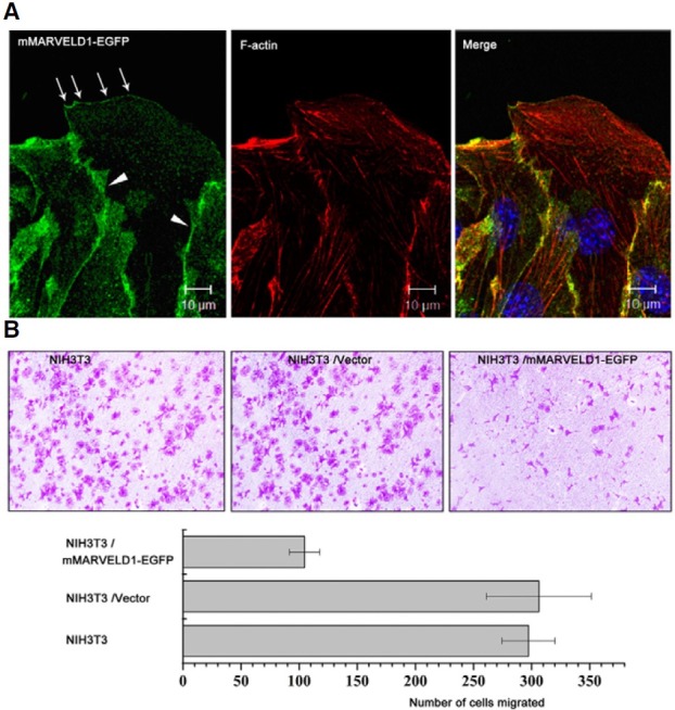
Fig. 6. Overexpressed mMARVELD1 localizes at the leading edge of migrating NIH3T3 cells and inhibits cell migration. (A) Localization of overexpressed mMARVELD1 in NIH3T3 cells during wound healing. Arrows indicate leading edge localization of mMARVELD1-EGFP, and arrowheads indicate cell junction localization of mMARVELD1-EGFP. (B) NIH3T3 cells expressing mMARVELD1- EGFP or control vector and untransfected cells were used for analysis of chemotactic motility in response to a serum gradient in a Transwell migration assay. Cells that traversed the membrane were visualized by crystal violet staining. Migrated cells in the Millicell plate were counted and statistically analyzed. Results are the average of three independent experiments (P < 0.05).

