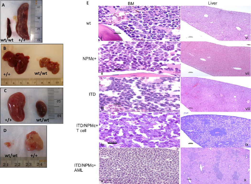
Figure 2. The leukemia that develops in ITD/NPMc+ mice is aggressive, infiltrating multiple organs.
Gross evaluation of representative leukemic ITD/NPMc+ mice, revealing involvement of the A) spleen, B) liver, C) kidneys, and in some cases the D) thymus. E) H&E stains of BM (i–v) and liver (vi–x) of representative mice of each genotype. Scale bars are as follows: i–v 10µm, vi–x 100µm.

