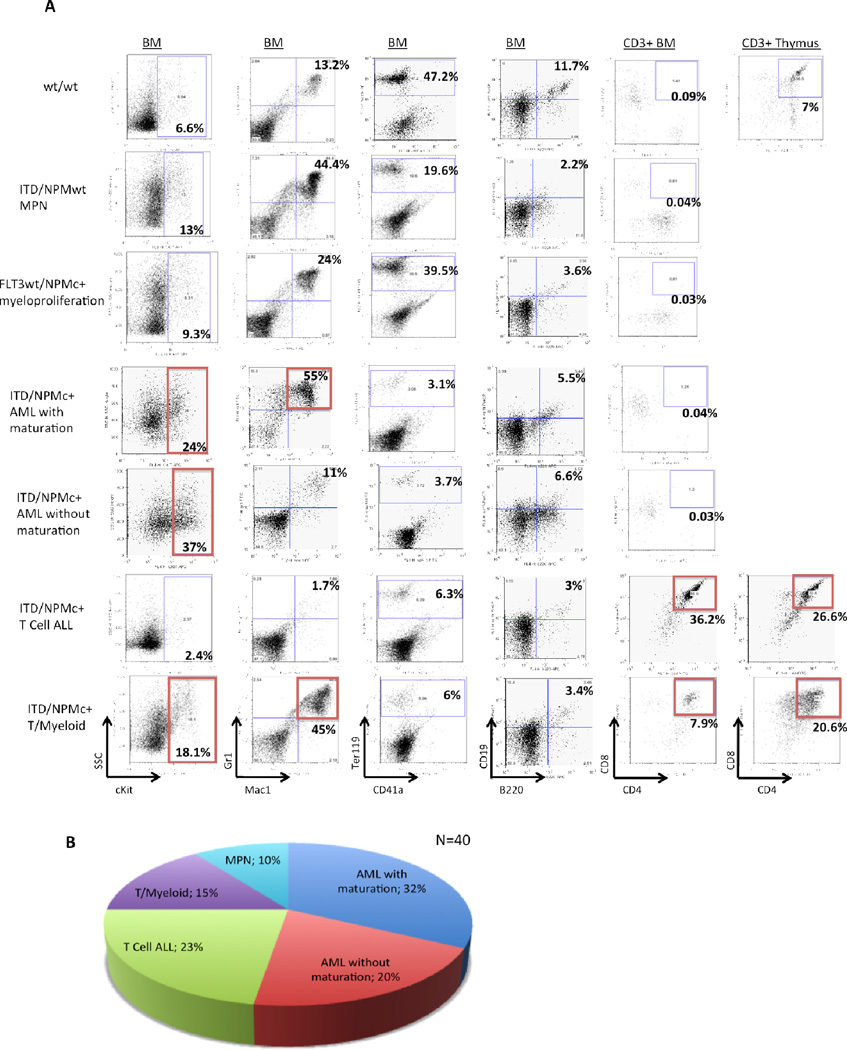
Figure 3. ITD/NPMc+ mice develop myeloid and lymphoid leukemias.
A) Flow cytometry plots demonstrating the characteristic phenotype of wt mice (row 1), ITD mice with myeloproliferative neoplasm (row 2), NPMc+ mice with myeloproliferation (row 3) and each of the 4 most common types of acute leukemia diagnosed in ITD/NPMc+ mice (rows 4–7). All the leukemic mice also have a paucity of normal maturing erythrocytes as demonstrated by decreased Ter119+ cells in the BM and maturing B lymphocytes indicated by decreased B220+/CD19+ cell in the BM. B) Disease distribution of the 40 fully characterized ITD/NPMc+ mice.

