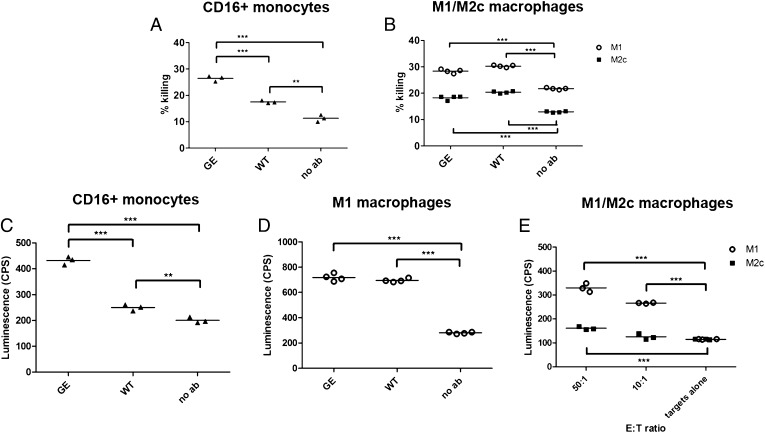
FIGURE 4.
Macrophage-mediated cytotoxicity. (A) A549 cells were incubated with isolated human intermediate, nonclassical (CD16+) monocytes (E:T ratio, 4:1) for 24 h in the presence of 1 μg/ml GA201 GE or WT Abs. (B) MKN45 cells were incubated with M1 or M2c macrophages (E:T ratio, 3:1) for 24 h in the presence of 1 μg/ml GA201 GE or cetuximab Abs. The percent killing was determined by setting the LDH released from target cells to 0% and LDH released by target cells lysed with 2% Triton X-100 to 100%. Triplicates and quadruplicates referring to one representative experiment out of three are shown. (C) 1833-PPOP233 cells were incubated with isolated human intermediate, nonclassical (CD16+) monocytes (E:T ratio, 30:1) for 8 h in the presence of 1 μg/ml GA201 GE or WT Abs. (D) 1833-PPOP233 cells were incubated with polarized human M1 macrophages (E:T ratio, 30:1) for 24 h in the presence of 1 μg/ml GA201 GE or WT Abs. (E) 1833-PPOP233 cells were incubated with polarized human M1 or M2c macrophages (E:T ratio, 10:1 or 50:1) for 24 h in the presence of 1 μg/ml GA201 GE. Triplicates and quadruplicates referring to one representative experiment out of three are shown. Statistical analysis, unpaired t test: *p < 0.05, **p < 0.01, ***p < 0.001.

