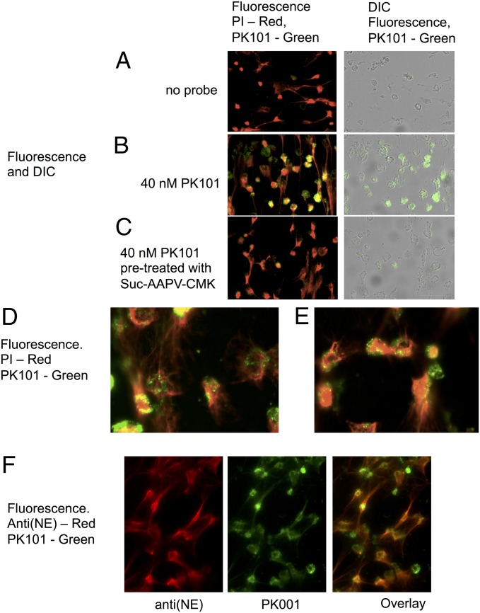
Fig. 5.
Imaging of neutrophils and NETs treated with PK101. NET formation was triggered with phorbol ester, followed by 20 min of treatment with 40 nM PK101. It was briefly washed, fixed, and incubated with fluorescent streptavidin to visualize the probe, and PI was used to visualize DNA. (A) No probe. (B) Probe added. (C) Samples treated with 10 mM MeOSuc-Ala-Ala-Pro-Val-CH2Cl for 30 min before addition of probe. (A–C) Imaged using a 20× objective; two overlaid images from the same experiment are seen in D and E with a 40× objective. Representative images of 4 experiments using 2 different donors. NET formation is visible as PI-stained strings and dispersed nuclear material, and PK101 labeling is visible as punctate staining associated with cellular material, but not with NETs. NETs treated with PK101 followed by anti(NE) antiserum and imaged using a 40× objective are shown in F. The total NE antigen parallels PI binding, demonstrating that NETS are decorate with NE, but the overlaid image demonstrates than most of the NE on NETs is not active but is concentrated in the cell-associated punctate structures.

