Fig. 3.
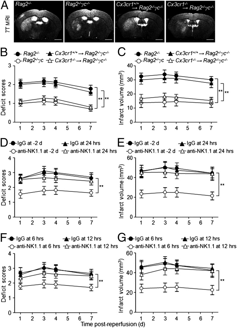
NK cells are associated with brain infarct volume. (A–C) Representative 7T MRI images (A) and quantification of neurological deficits (B) and infarct volumes (C) in MCAO mice with NK cells (Rag2−/−) vs. without NK cells (Rag2−/−γc−/−), as well as more (Cx3cr1+/+ NK→Rag2−/−γc−/−) vs. less (Cx3cr1−/− NK→Rag2−/−γc−/−) NK cells in the brain. Rag2−/−γc−/− MCAO mice had relatively mild neurological deficits and smaller infarct volumes than Rag2−/− MCAO mice. Reconstitution of Cx3cr1+/+ but not Cx3cr1−/− NK cells restored the ischemic lesions in Rag2−/−γc−/− mice. Data generated from 15 mice per group. **P < 0.01. (Scale bars, 1 mm.) (D–G) Determination of the time window in which NK cells exert detrimental effects in stroke. WT mice were treated with anti-NK1.1 mAb or isotype control IgG Ab 2 d before MCAO or at 6, 12, and 24 h after reperfusion, respectively. Treatment regimen and efficiency of cell depletion are described elsewhere (16, 30). Neurological deficits (D and F) were assessed, and infarct volumes (E and G) were determined by MRI in conjunction with TTC staining. Attenuation was more pronounced when NK cells were depleted preceding MCAO or within the first 12 h after MCAO. n = 8 per group. **P < 0.01.

