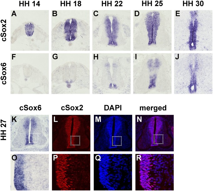
Fig. 2.
Expression of Sox2 and Sox6 in the developing chick neural tube. RNA in situ hybridization was performed for Sox2 (A–E) or Sox6 (F–J) on adjacent transverse sections at the forelimb levels of the chicken embryos at HH stages 14 (A and F), 18 (B and G), 22 (C and H), 25 (D and I), and 30 (E and J). Coexpression of Sox6 and Sox2 at the cellular level was examined by staining for Sox6 by RNA in situ hybridization (K and O) and for Sox2 by immunohistochemistry (L and P). Sox2 immunostained sections were DAPI-counterstained (M and Q), and merged images (N and R) show that virtually all cells in the ventricular zone are positive for Sox2. Boxed areas in K, L, M, and N are presented in enlarged forms in O, P, Q, and R, respectively.

