Fig. 1.
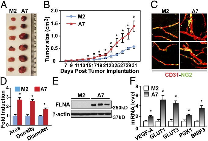
FLNA-expressing tumor xenografts grow faster and show increased angiogenesis and HIF-1α activity. (A) Gross view of tumor xenografts 31 d after injection of M2 or A7 cells into SCID mice. (B) FLNA-expressing tumor xenografts (A7) grow faster than tumor xenografts that do not express FLNA (M2). (C and D) FLNA-expressing tumor xenografts have increased angiogenesis (n = 6). (C) Tumor vasculature was studied with a whole-mount method using anti-CD31 and anti-NG2 antibodies to label the endothelial cells (red) and pericytes (green). (D) Tumor vessel area, density, and vessel diameter were evaluated (n = 6). (E) Expression of FLNA and β-actin in xenograft tumor samples was detected by immunoblotting. (F) FLNA-expressing tumor xenografts (A7) express higher levels of HIF-target genes (n = 3). Data are shown as mean ± SD. *P < 0.05 compared with corresponding samples of M2-derived tumor xenografts.

