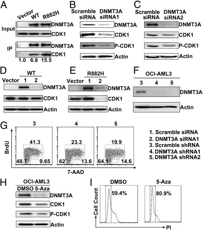
Fig. 5.
Protein–protein and functional interaction between the DNMT3A mutant and CDK1. (A) Interaction of DNMT3A with CDK1. Lysates from 293T cells transfected with plasmids encoding Flag-DNMT3A were immunoprecipitated using anti-Flag and analyzed by Western blot using the indicated antibodies. (B and C) Lysates from OCI-AML3 cells transfected with two siRNAs (siRNA1 and 2) against different DNMT3A regions were Western blotted with the indicated antibodies. (D and E) Analysis of lysates from NIH3T3 cells stably expressing DNMT3A WT or the R882H mutant. These cells were transfected with scramble siRNA or DNMT3A siRNA1, and their lysates were Western blotted with antibodies against the indicated proteins. Vector-transfected cells were used to estimate the endogenous CDK1 level compared with levels in cells transfected with WT or mutant DNMT3A. (F) OCI-AML3 cells were infected with lentiviruses expressing scramble shRNA (control) or two shRNAs (shRNA 1 and 2) targeting different DNMT3A regions. (G) OCI-AML3 cells infected with lentiviruses expressing control or two different DNMT3A shRNAs were labeled with BrdU and 7-AAD for flow cytometry analysis. (H and I) Western blot analysis of indicated proteins (H) and propidium iodide (PI) staining flow cytometry analysis (I) in OCI-AML3 cells treated by DMSO or 5-Aza (0.8 μM) for 48 h.

