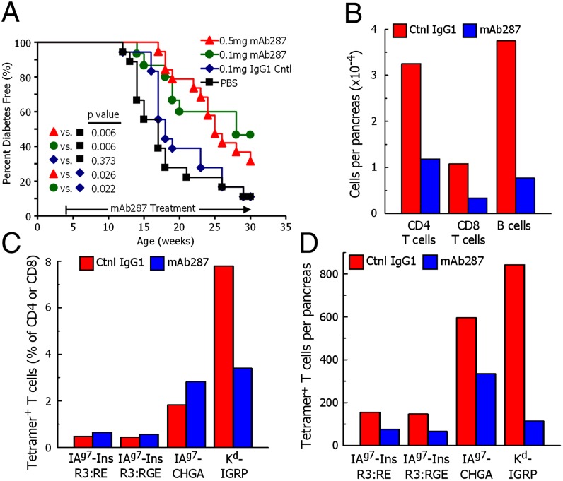
Fig. 3.
Monoclonal antibody mAb287 delays diabetes and islet lymphocyte infiltration in NOD mice. (A) Groups of 4-wk-old female NOD mice were treated weekly with PBS (n = 18; black squares), 0.1 mg of mouse IgG1 (n = 18; blue diamonds), 0.1 mg of mAb287 (n = 15; green circles), or 0.5 mg of mAb287 (n = 18; red triangles), and followed up to 30 wk. Diabetes was diagnosed as described in Materials and Methods. The percentages of remaining diabetes free of each group are shown. P values were determined by using χ2 log rank test. (B–D) Groups of eight mice were treated weekly from age 4–11 wk with 0.5 mg of control IgG1 (red bars) or mAb287 (blue bars) at which time pancreatic islets were pooled in each group. A cell suspension was prepared and analyzed by flow cytometry. (B) The average number of live CD4 T cells, CD8 T cells, and 220+ B cells per pancreas were estimated in each sample. (C) The percentages of CD4 T cells specifically binding the IAg7–R3:RE, IAg7-R3:RGE or IAg7-ChgA tetramers, or CD8 T cells specifically binding the Kd-IGRP tetramer were determined as describe in Fig. S6. (D) Average number of islet-infiltrating tetramer-positive cells per pancreas were calculated from the data in B and C. In a second experiment, analysis with the IAg7 tetramers was repeated with similar results by using islets pooled from five mice in each group.

