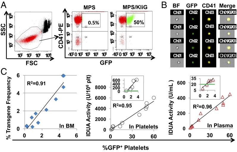
Fig. 2.
In vivo expression of both GFP and lysosomal IDUA in platelets of MPS I chimeras after transduction with erythroid/MK-specific LV. Lin− cells from MPS I mice were transduced with LV-KIiG and transplanted into lethally irradiated MPS I mice (MPS/KIiG). MPS/KIiG mice with various gene dosages were generated by 2° transplantation using a serial dilution of BM from 1° MPS/KIiG with MPS marrow 5 mo after BM transplantation. (A) Representative GFP expression in platelets is shown by FACS plots after staining with CD41-PE antibody. FSC, forward light scatter; SSC, side light scatter. (B) Representative GFP+ platelets are viewed by ImageStream analysis. (C) Correlation of GFP expression in platelets with gene transfer efficiency (GT) in BM cells as determined by real-time qPCR, IDUA enzyme activities in isolated platelets and in plasma from MPS/KIiG mice. The Insets show IDUA levels in mice at ≤4% GT, with green lines indicating WT levels. Each symbol represents data from an individual animal.

