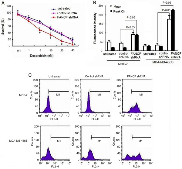
Figure 7. Effects of FANCF-specific shRNA on Dox sensitivity of MCF-7 and MDA-MB-435S cells. A, Cells were treated with various concentrations of Dox. Cell viability was determined with a CCK-8 kit. The percentage of viable cells was determined by the ratio of viable cells treated with FANCF shRNA or control shRNA to that with no treatment. B, Median fluorescence intensity was measured indicating the relative amount of Dox accumulation. C, Effects of FANCF shRNA on cellular Dox accumulation. Cells were transfected with FANCF shRNA or control shRNA for 48 h following a 24-h incubation with 10 nM Dox. Cellular uptake of Dox was measured by fluorescence-activated cell sorting. Data are reported as means±SD of three independent experiments in triplicate. *P<0.05, ANOVA followed by the post hoc test.

