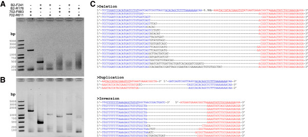
Figure 3.

Detection of large chromosomal structural variation. (A and B) PCR was performed using primer set to detect chromosomal deletion (lane 4), duplication (lane 5), and inversions (lane 6 and 7) from genomic DNA of un-injected embryos (A) and injected embryos (B). For comparison, primer sets to amplify the flanking regions of TALEN-702 site (lane 2) and TALEN-B2 site (lane 3) were also used. M represents DNA ladders, the letters on the left indicate length of each band. (C) Sequences of the PCR products. The wild-type sequence is shown at the top with the full TALEN-B2 site in blue and the TALEN-702 site in red. The TALEN recognition sites are underlined, and deletions are indicated by dashed lines.
