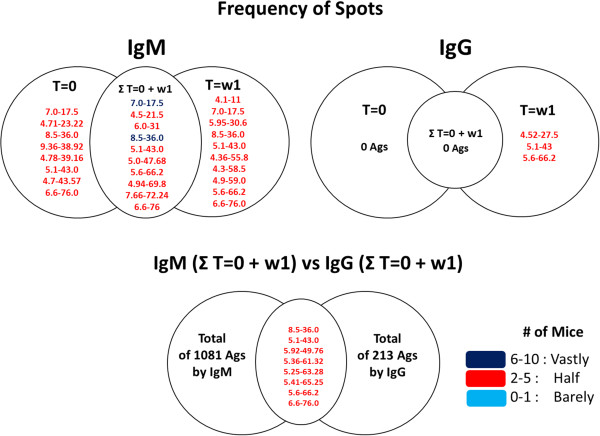
Figure 3.

Frequency of spots found in each 2D-immunoblot images of female mice in T = 0 and w1 of response mediated by IgG and IgM. Figure shows the isoelectric point and molecular weigth of spots that are shared not only among images of each time (T = 0 and T = w1), but also among all female mice. Spots in red color are recognized "Half" and spots in blue heavy color are recognized "Vastly" by mice. Spots are recognized by 0 to 1 mice were categorized as "Barely", by 2 to 5 mice as "Half" and by 6 to 10 mice as "Vastly". Ags = antigens.
