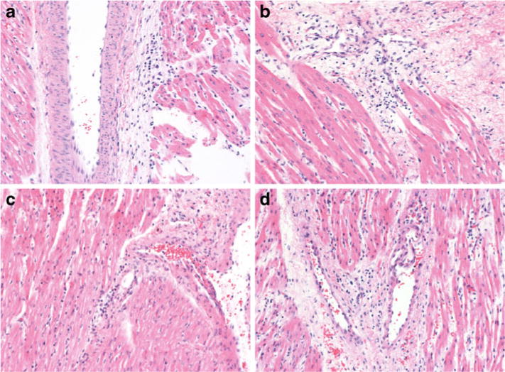
Fig. 5.

Representative samples of slides used for histological evaluation of the left and right ventricles in the control and insulin groups. a and b Hematoxylin and eosin stain of left ventricle from control group and insulin group, respectively. c and d Hematoxylin and eosin stain of right ventricle from control group and insulin group, respectively. Slide thickness is 5 μm. Magnification is 200×.
