
Figure 3. Impaired blood flow recovery and reduced capillary density in both male and female ARKO mice after ischemia.
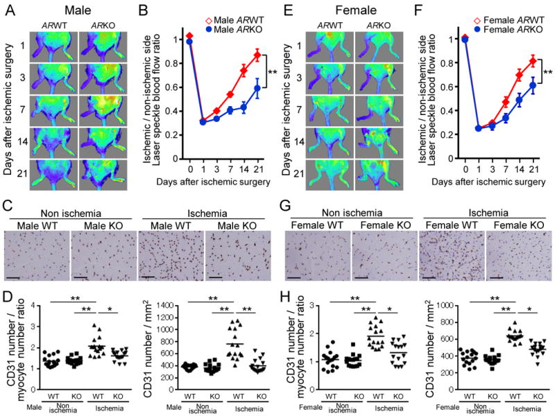
(A and E) Representative laser speckle blood flow (LSBF) images of the ischemic hind limb in WT and ARKO mice during the experimental period. A low perfusion signal is indicated as dark blue and according to increase in perfusion signals, the colors are indicated as green, yellow and red in series. (A) Male WT mice (n=17) and male ARKO mice (surviving hind limbs, n=8). (E) Female WT mice (surviving hind limbs, n=16) and female ARKO mice (surviving hind limbs, n=7). (B and F) Cumulative results are shown as ratio of blood flow in the ischemic limb to that in the nonischemic limb at each time point (B: male mice, F: female mice). **P<0.01 using a linear mixed effects regression analysis. Error bars represent ± SEM. (C and G) CD31 immunohistochemical staining to determine capillaries in ischemic and nonischemic thigh adductor muscles in WT and ARKO mice at day 21 after surgery (C: male mice, G: female mice). Scale bar indicates 100 μm. (D and H) Quantification of capillary density is expressed as CD31-positive number per myocyte number or per square millimeter in WT and ARKO mice (D: male mice, H: female mice). n=16 in each group. *P<0.05, **P<0.01 using Dunn's test following the Kruskal-Wallis test. Bars represent mean values in each group.