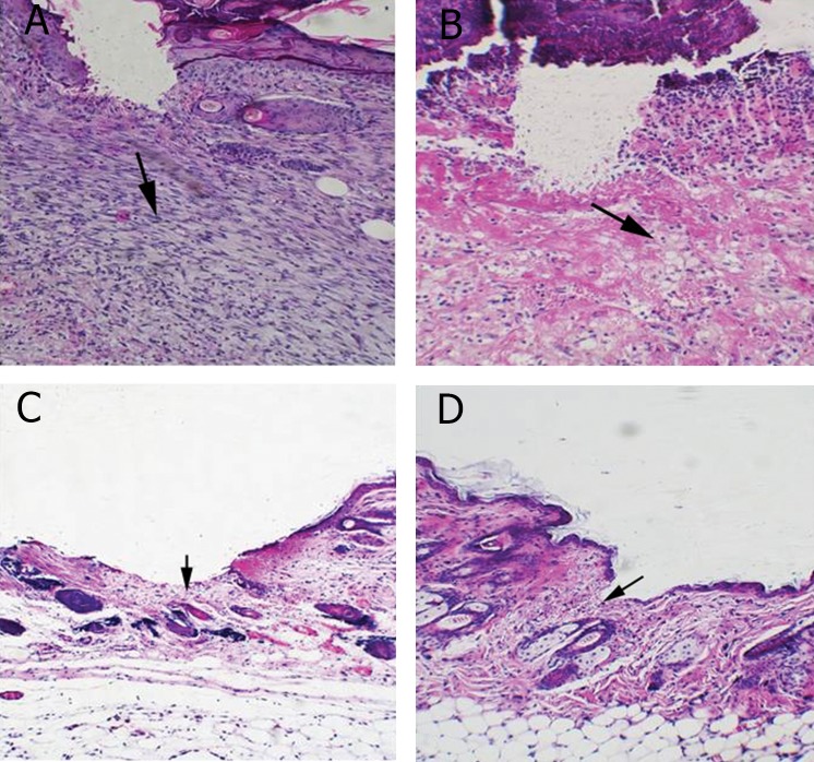
Fig 2.
Histological analysis of H&E stained skin tissues from wounds on 3 and 7 days post incision. A. Cell infiltration increased in the wound site on day 3 post-incision in control group (arrow). B. In Oleuropein treated mice infiltrated cells were less than control group (arrow). C. Skin tissues from wound on day 7 post-incision in control group is shown, please note that epithelialization was not completed (arrow). D. Wound tissues from Oleuropein treated mice showed an advanced re-epithelialization (arrow) and dermal regeneration with significant decrease in cell infiltration compared to control group (Magnification ×200).

