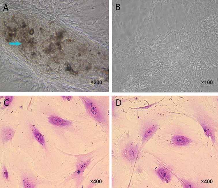
Fig 1.
Characteristics of osteoblast and osteoclast derived from mouse dental pulp stem cells. Morphology of stem cells after 21 days exposed to osteoblast induction medium (A). The morphology of mouse dental pulp cell before induction (B). The blue arrows show calcium salt with a dark brown color, whereas the control group that was not exposed to osteoblast induction did not show any calcium nodule. In osteoclast induction medium, the morphology of mouse dental pulp cell after induction (C). Morphology of stem cells before exposing to osteoclast induction (D). Osteoclast cells were stained by May-Grunwald-Giemsa. The staining indicated multinucleated cells were found in dental pulp stem cells cultured in both medium, i.e., osteoclast differentiation medium and complete medium (control).

