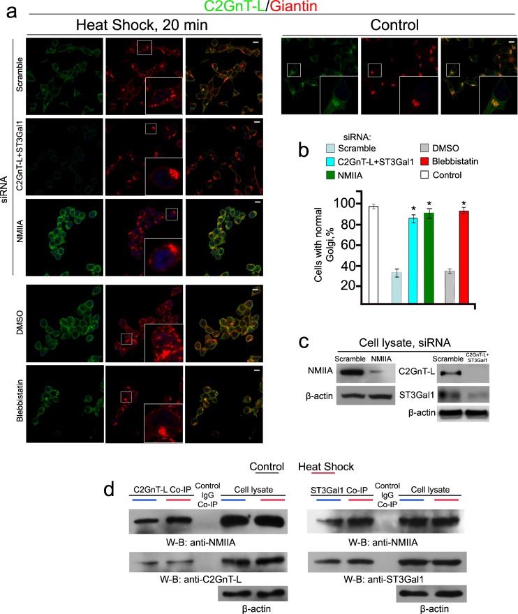
Fig. 4.
Depletion of C2GnT-L and ST3Gal1 in LNCaP cells delays HS-induced Golgi disorganization. a Confocal immunofluorescence images of C2GnT-L and giantin in cells treated with scramble, NMIIA, or C2GnT-L + ST3Gal1 siRNAs for 72 h or 35 μM blebbistatin for 1 h and appropriate amount of DMSO followed by HS at 45 °C for 20 min. White boxes in the images indicate Golgi (giantin) areas enlarged in the inset. b Percentage of the cells with fragmented Golgi in cells described in a: KD of both C2GnT-L and ST3Gal1 delayed Golgi disorganization similar to NMIIA KD or inhibition. The data are expressed as mean ± SEM; Asterisk, p < 0.001. c NMIIA, C2GnT-L, and ST3Gal1 western blot of the lysates of cells treated with scramble, NMIIA (MYH9) or ST3GAL1 + GCNT1 (C2GnT-L) siRNAs; beta-actin was used as a loading control. d NMIIA and C2GnT-L/ST3Gal1 western blots of complexes pulled down with anti-C2GnT-L or ST3Gal1 Ab from the lysates of cells immediately after HS. Cell lysates containing same amounts of proteins were used for Co-IP. The results shown are representative of three independent experiments. All confocal images were acquired with same imaging parameters; bars, 10 μm

