
Figure 6. Merger of red (band3) and green (spectrin) channels after examination in a confocal laser scanning microscope.
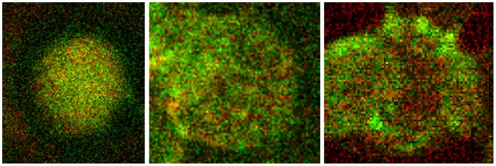
Double stained erythrocytes with antibodies against band3 (red) and spectrin (green, details from Figures 1 , 2 , 3 ). Left: erythrocyte suspended in autologous plasma, middle: erythrocyte suspended in a Iodixanol/plasma-mixture (30% v/v), right: erythrocyte suspended in a Iopromide/plasma-mixture (30% v/v; primary magnification 1∶63; zoom factor 1∶10).