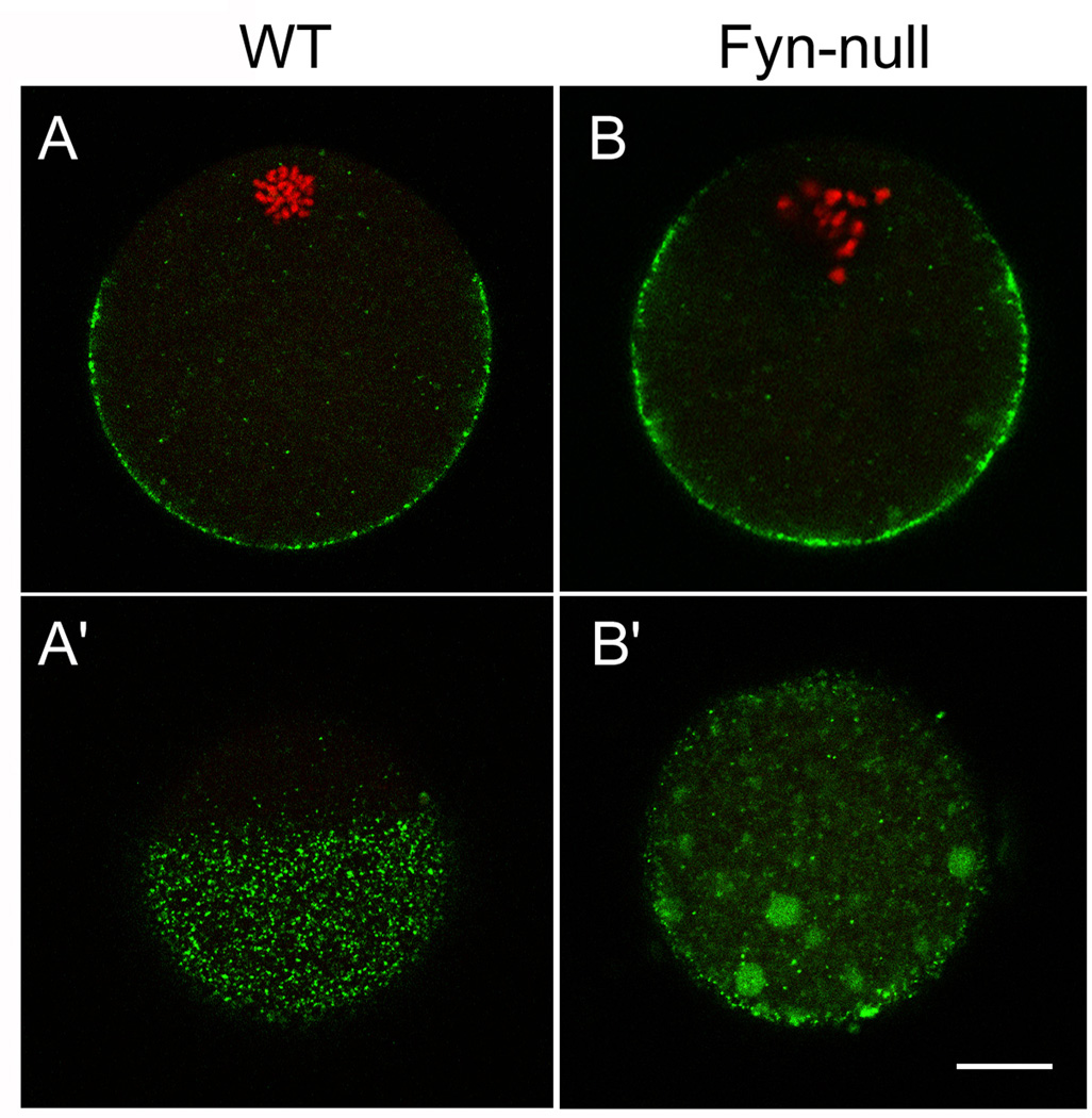
Figure 4.
Distribution of cortical granules in Fyn-null oocytes. Cortical granules were detected in (a, a’) wild-type (WT) and (b, b’) Fyn-null oocytes by labeling oocytes with lectin from Lens culinaris (LCA)–fluorescein isothiocyanate (FITC), with FITC fluorescence appearing green. The DNA was labeled with ethidium homodimer and appears red in this figure. Photographs taken through the oocyte equator demonstrate the extent of the cortical granule-free zone in (a) WT oocytes (arrows), which was reduced in Fyn-null oocytes (b). Images taken tangentially through the oocyte cortex (a’, b’) demonstrate the distribution pattern of cortical granules within the cortical cytoplasm. Fyn-null oocytes displayed irregular clusters of cortical granules that were rarely seen in control oocytes. Scale bar < 10 mm.

