Figure 5.
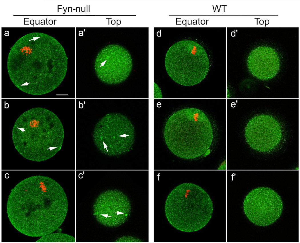
Distribution of Type 1 inositol 1,4,5-trisphosphate receptor (IP3R1) in Fyn-null oocytes. Immunofluorescence detection of IP3R1 in Fyn-null MII oocytes (a–c, a’ –c’) and wild-type (WT) MII oocytes (d–f, d ‘ –f ‘) that were stained with rabbit anti-IP3R1 antibody, followed by an Alexa fluor 488-labelled secondary antibody (green). Optical sections taken through the equator of the oocyte demonstrate the distribution of IP3 receptors in the oocyte cortex and central cytoplasm (a–f). Tangential optical sections (a’ –f ‘) reveal the distribution in the cortex just below the oocyte surface. DNA was stained by ethidium homodimer-2 (red). Arrows indicate dense accumulation of IP3R1 clusters in Fyn-null oocytes. Scale bar = 10 mm.

