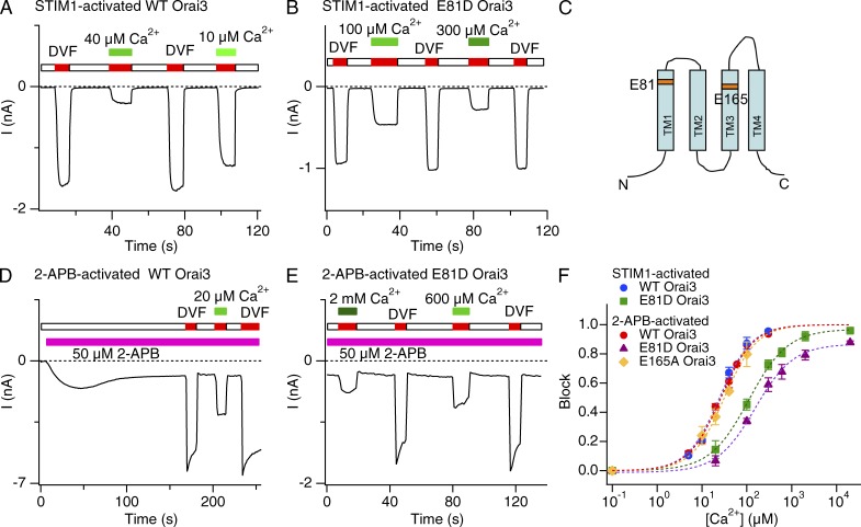
Figure 2.
Extracellular Ca2+ blocks STIM1- and 2-APB–activated Orai3 Na+ currents with similar sensitivity. (A, B, D, and E) Inhibition of STIM1- or 2-APB–activated Orai3 Na+ currents by Ca2+o. In each case, the cell was voltage clamped to a constant potential of −100 mV and 200-ms sweeps were collected at 2 kHz. The mean current during each sweep is plotted against time. (C) Predicted topology of a single Orai3 subunit with the acidic residues (TM1 and TM3) highlighted. (F) Dose–response relationships of Ca2+ blockade. Ca2+ blockade of Na+ currents through 2-APB–activated WT and E165A Orai3 channels occurred with a similar affinity as STIM1-activated Orai3 channels. E81D substitution increases the Ki of block in both STIM1- and 2-APB–activated modes (also see Table 1). Block was quantified by measuring the Na+ current immediately after application of a DVF solution supplemented with the indicated [Ca2+]o. Each dashed line is a least-squares fit of the Hill equation block = max/[1 + (Ki/[Ca])n], where max is the predicted maximal blockade at saturating Ca2+ concentrations. Error bars represent SEM.

