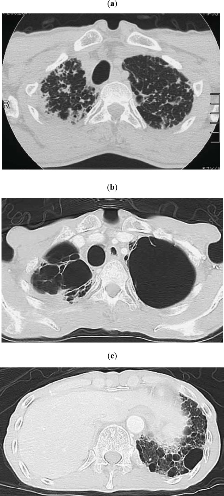
Fig. (4).
(Same patient as in Fig. 3) (a) Chest CT showing subpleural nodular or reticular opacities in the lung parenchyma at the apex. Interlobular septal thickening was associated (taken 7 years after the image shown in Fig. 3a). (b) Chest CT taken 2.8 years after the image shown in 4a. Multiple bullae and large cysts appeared in the upper lung fields. (c) Chest CT taken 2.8 years after the image shown in 4a. Multiple fibrocystic changes appear in the lower lobes.

