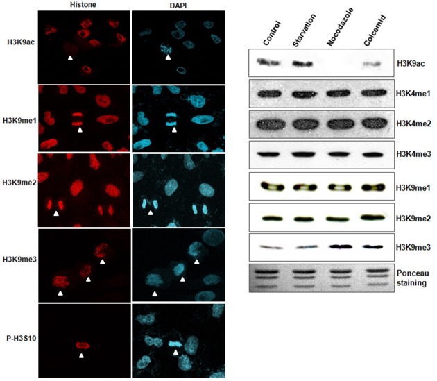
Fig. 1. Changes of histone modifications during cell cycle progression. (A) Histone modifications (red) were determined by immunostaining in culturing A549 cells. DNA was DAPI-stained for assumptions of mitotic stage. Acetylation of H3K9 (H3K9ac) was reduced in the mitotic cells, but trimethyation of H3K9 (H3K9me3) was increased in the mitotic cells. However, monomethylation of H3K9 (H3K9me1) and dimethylation of H3K9 (H3K9me2) were not changed under identical conditions. P-H3S10 is shown as a mitotic control. White arrow head; mitotic cells. (B) Western blot analyses were conducted using synchronized HeLa cells. H3K9me3 and H3K9ac expressions were altered in the nocodazole and colcemid treatment groups, but not in the serum star-vation group.

