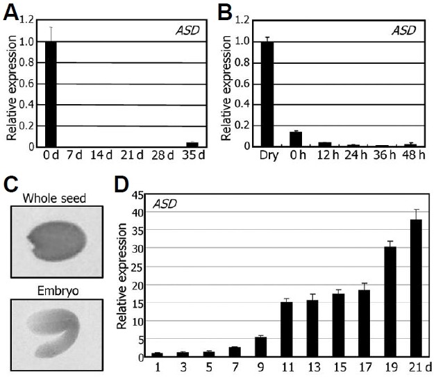
Fig. 2. Temporal and tissue-specific expression patterns of the ASD gene. In (A), (B), and (D), transcript levels were examined by using qRT-PCR. Biological triplicates were averaged. Bars represent standard error of the mean. (A) Temporal expression pattern. Total RNA was extracted from seeds or whole seedlings harvested at the indicated time points. d, days after germination. (B) Expression pattern in germinating seeds. Total RNA was extracted from the seeds harvested at the indicated time points. h, hours after cold imbibition. (C) Distribution of GUS activity in seeds. The pASD-GUS fusion construct, in which the GUS-coding sequence was transcrip-tionally fused to the ASD gene promoter sequence covering an approximately 1-kb region upstream of the transcription start site, was transformed into Col-0 plants. GUS activities were detected exclusively in the embryo. (D) Expression pattern during silique development. Total RNA was extracted from the siliques harvested at the indicated time points. Silique development covered from anthesis (0 DAP) to dry seed stage (21 DAP). d, days after pollina-tion (DAP).

