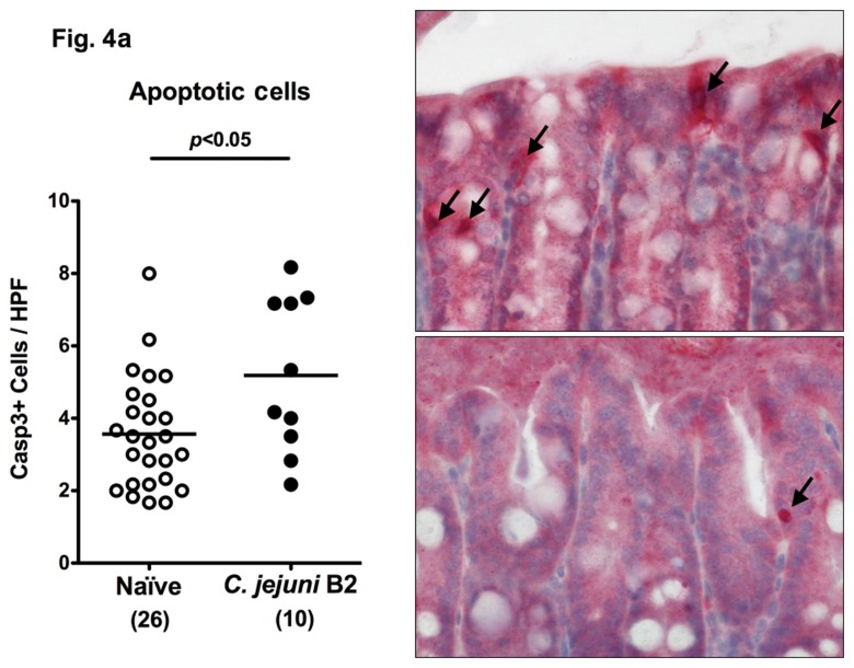
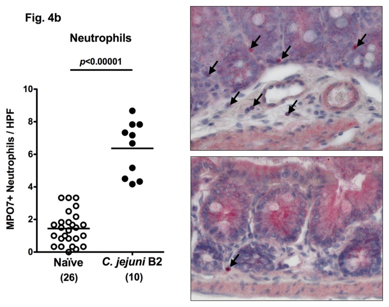
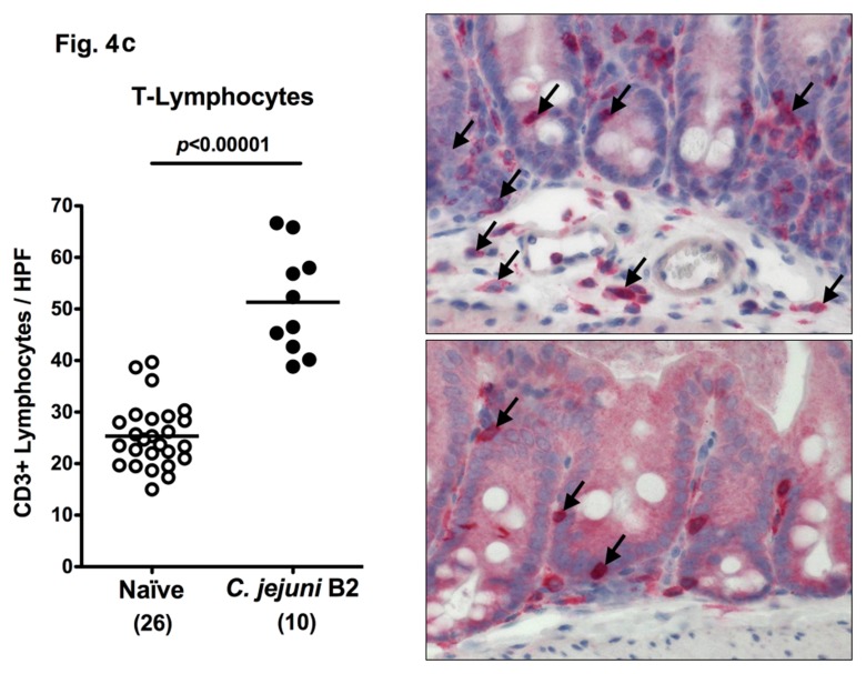
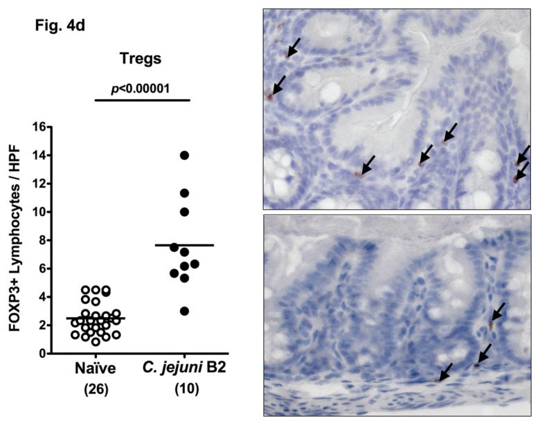
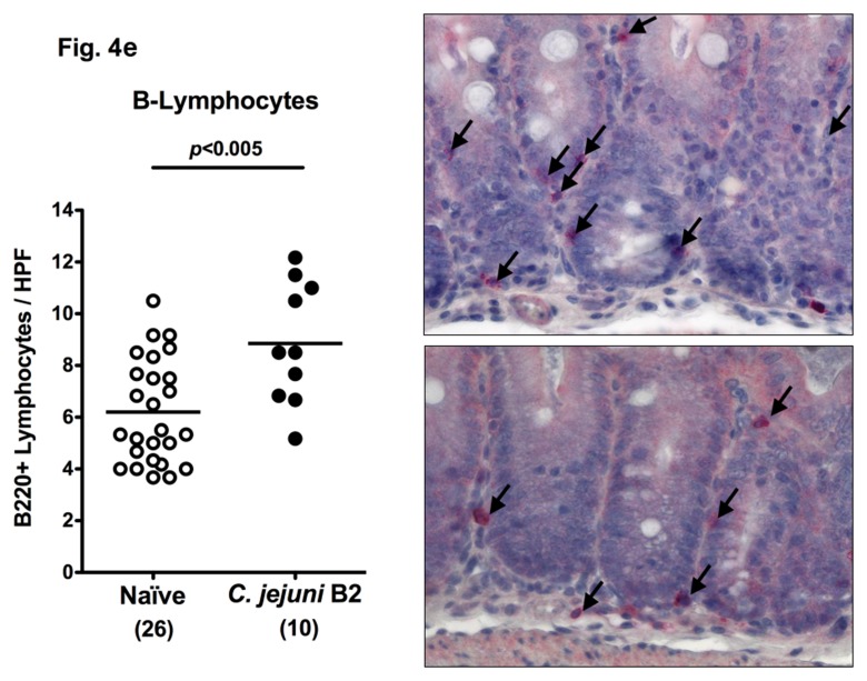
Fig. 4.
Immune cell response in colon sections of C. jejuni B2 infected infant mice at day 103 p.i. Infant mice 3 weeks of age were infected with C. jejuni B2 right after weaning, and paraffin sections of colon samples obtained at day 103 p.i. (solid circles). Uninfected infant mice (naïve, open circles) served as negative controls. The average numbers of apoptotic cells (positive for caspase-3, panel A), neutrophilic granulocytes (neutrophils, positive for MPO-7, panel B), T-lymphocytes (positive for CD3, panel C), regulatory T-cells (positive for FOXP3, Tregs, panel D), and B-lymphocytes (positive for B220, panel E) from at least six high power fields (HPF, ×400 magnification) per animal were determined microscopically in immunohistochemically stained colon sections (left panel). Representative photomicrographs of positively stained cells in colon section (right panel; solid arrows) at day 103 p.i. (upper panel) versus naïve controls (lower panel) from three independent experiments are shown (HPF, ×400 magnification). Data were pooled from at least three independent experiments. Numbers of animals are given in parentheses. Means (black bars) and levels of significance (P-values) as determined by the Student’s t-test are indicated

