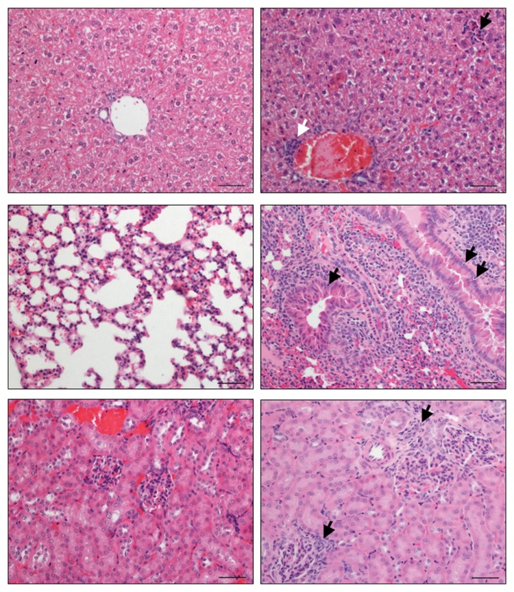
Fig. 5.
Long-term extra-intestinal inflammatory responses in C. jejuni B2 infected infant mice. Infant mice 3 weeks of age were infected with C. jejuni B2 right after weaning. Paraffin sections of ex vivo biopsies taken from livers (upper panel), lungs (middle panel), and kidneys (lower panel) at day 103 p.i. (right panel) were HE-stained as described in the section Materials and methods. Uninfected infant mice (left panel) served as negative controls. Representative photomicrographs from three independent experiments are shown (×200 magnification, scale bar 50 μm). Lymphocytes infiltrating the periportal field (solid white arrow, upper right), the liver parenchyma (solid black arrow, upper right), and the glomerular kidney tubuli (solid black arrows, lower right) are indicated. Solid black arrows in the lung (middle right) point toward apoptotic cells

