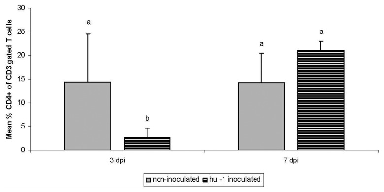
Fig. 2.
Comparison of the relative percent of CD4+ T cells of intraepithelial lymphocytes (IELs) of the jejunum of a non-inoculated and a group, inoculated with a human (hu) C. jejuni strain at 3 and 7 days post inoculation. n = six animals/day and group representative for the four conducted experiments. IELs were isolated and processed for flow cytometric analysis using monoclonal antibodies for CD3+ and CD4+ T cells (Southern Biotechn., USA). Presented are the %CD4+ T cells within the CD3+ IEL. Different letters indicate significant differences between groups at indicated time-points (p ≤ 0.05 Kruskal–Wallis test).

