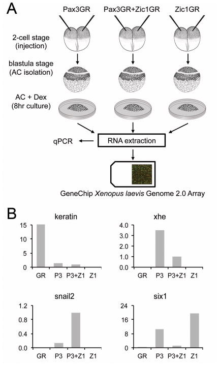
Figure 1. Strategy to isolate Pax3-Zic1 targets.
(A) Procedures used to identify Pax3-Zic1 targets in the developing NC. Xenopus embryos were injected at the 2-cell stage with mRNA encoding GR (not shown), Pax3GR and Zic1GR (250 pg each), alone or in combination. At the blastula stage (stage 9), animal cap explants (AC) were dissected and cultured for 8 hours in the presence of dexamethasone. RNA were extracted from each sample, analyzed by qPCR and subsequently used to screen a GeneChip Xenopus laevis Genome 2.0 Array (Affymetrix). (B) RNA extracted from GR, Pax3GR, Zic1GR, or Pax3GR+Zic1GR injected animal cap explants, were analyzed by qPCR for the expression of various marker genes, keratin (epidermis), xhe (HE), snail2 (NC), and six1 (PE) to confirm that the expected pattern of gene expression was observed in each injection group. The relative expression levels have been normalized to ef1α. P3, Pax3GR; Z1, Zic1GR.

