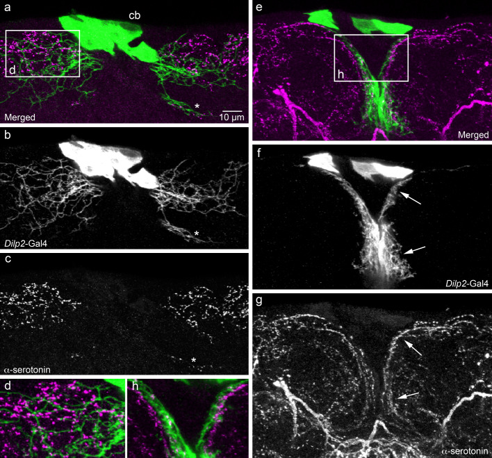
Fig. 3.
Relationships between processes of IPCs and serotonin-immunoreactive neurons in the adult brain. We utilized the Dilp2-Gal4 to drive GFP in IPCs (green) and a mouse monoclonal antibody to serotonin (magenta) to visualize relationships between the two neuron types in the pars intercerebralis (shown in frontal view; dorsal up). a–c IPCs at the level with wide lateral branches dorsally. These IPC branches superimpose those of varicose serotonin immunoreactive ones (even the small set of branches more ventrally, at asterisk). d The framed area in (a) is shown at higher magnification. e–g The IPCs at the level of the median short branches (other specimen). Again, the branches of the two neuron types superimpose (e.g., at arrows). h The framed area in (e) is shown at higher magnification

