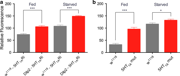
Fig. 4.
Knockdown of 5-HT1A leads to an increase in DILP-immunolabeling in insulin-producing cells (IPCs). a Relative DILP immunofluorescence in IPCs in fed and starved flies with and without 5-HT1A receptor knockdown (5-HT1ARi) in IPCs (Dilp2-Gal4/UAS-5-HT 1A-RNAi). The DILP2 antiserum used is likely to cross react with DILP2, 3 and 5. Control flies (w1118-5-HT1A) display significantly lower levels of DILP-immunofluorescence than the flies with diminished 5-HT1A (Dilp2-5-HT1ARi), both in fed flies (***p < 0.001; One-way ANOVA with Tukey’s comparison) and after starvation (***p < 0.001). Also, the increases of fluorescence in controls (gray bars) and knockdown flies (red bars) when comparing fed and starved flies are significant (p < 0.001 for both). For each genotype and condition, IPCs of 5–7 brains were investigated. b Relative DILP immunofluorescence in IPCs in fed and starved mutant (5-HT1A mut) and wild type (w1118) flies. Wild-type flies display significantly lower levels of DILP-immunofluorescence than the 5-HT1A mutant flies, both in fed flies (***p < 0.001) and starved ones (*p < 0.05). Again, the increases in immunolabeling in fed and starved controls and fed and starved mutants are significant (p < 0.001 for both). IPCs of 7–10 brains of each genotype and condition were investigated

