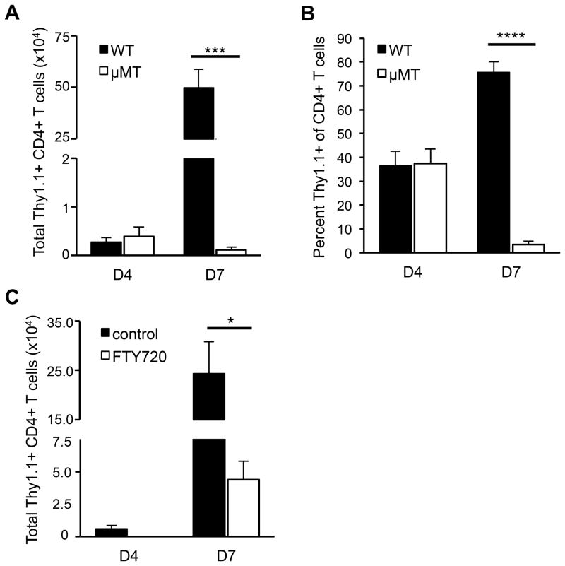
Figure 3. MOG-specific T cells fail to accumulate in the CNS in the absence of B cells.
Thy1.1+ T cells from MOG-immunized donors were activated in vitro with MOG97-114 and transferred into Thy1.2 WT or μMT recipients. CNS mononuclear cells were isolated on days 4 and 7 post-transfer and analyzed for (A) the total number of donor CD4+ Thy1.1+ T cells and (B) percent of Thy1.1+ donor cells among total CD4+ T cells (means ± SEM, n ≥ 5 mice per group). (C) FTY720 or vehicle was injected into WT mice daily starting on day 4 post-transfer of MOG-specific T cells. CNS cells were isolated from recipients prior to treatment on day 4 post-transfer and on day 7 from both treated and control recipients. The total number of CD4+ Thy1.1+ donor T cells in the CNS was determined by flow cytometry (means ± SEM, n ≥ 4 mice per group). (AC) Significant differences between means are indicated; *p < 0.05; ***p < 0.001; ****p < 0.0001, Student’s t test. Results are representative of at least 3 independent experiments.

