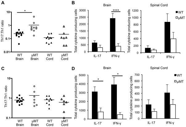
Figure 7. B cell-deficient recipients that developed EAE exhibit higher Th17:Th1 ratios in the brain compared to wild-type recipients.
EAE was induced by adoptive transfer of Th1- or Th17-skewed cells into C3HeB/Fej WT or μMT recipients. At EAE onset, CNS cells were isolated from the brain and spinal cord and plated in ELISPOT assays to detect IL-17- or IFN-γ-producing cells. Numbers of antigen-specific spots were determined by comparing spots in wells with and without MOG97-114. (A and B) Mice received Th1-skewed cells. (C and D) Mice received Th17-skewed cells. (A and C) The Th17:Th1 ratio is shown, calculated from the number of IL-17/IFN-γ producing cells in each culture. Each data point represents a single mouse. Data are pooled from at least 3 independent experiments. *p < 0.05; ***p < 0.001; ****p < 0.0001, Student’s t test.

