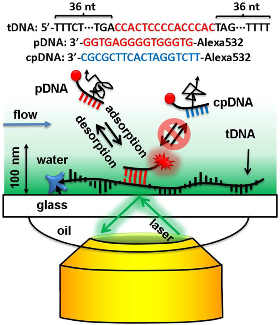
Figure 1.
Scheme of super-resolution imaging of immobilized target tDNA, with a fluorescently labeled probe pDNA, using mbPAINT and a TIRF microscope. Only the molecules within the evanescent field of the excitation light were excited and only those localized were observed. The negative control probe cpDNA, does not bind to the tDNA, so it diffuses freely in the solution and cannot be observed. Drawing not to scale.

