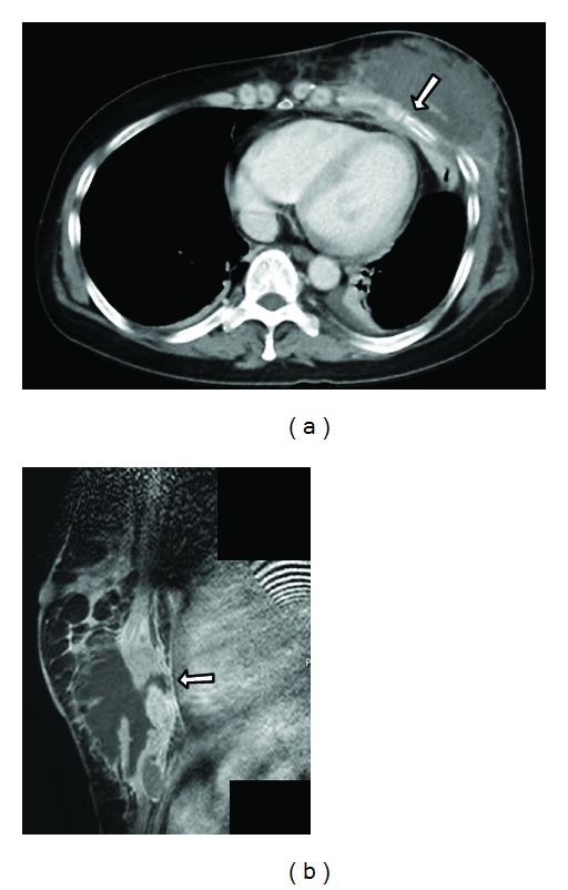
Figure 3.

(a) Computed tomography (CT) showing a large mass in the lower region of the left breast. The fifth rib under the mass is fractured (arrow). (b) T1-weighted magnetic resonance imaging reveals contrast enhancement from the border of the mass to the pleura (arrow).
