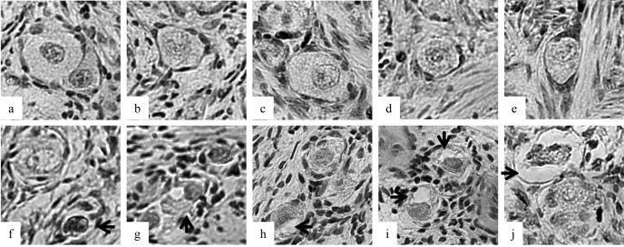
Fig. 1.
Morphologies of porcine follicles after vitrification. a–e: Follicles were categorized as normal after vitrification between the graphite sheets. f–j: Follicles were categorized as damaged after vitrification on the copper sheet. Each arrow shows a pyknotic oocyte nucleus (f), shrunken ooplasm (g–j) and disorganized granulose cells (j).

