Fig. 1.
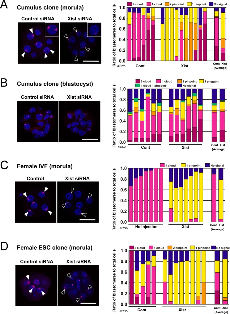
RNA-FISH analyses of Xist in cumulus cell-derived cloned embryos (A and B), female IVF embryos (C), and female ES cell-derived embryos (D). The right panels show the ratios of blastomeres classified according to the number of cloud or pinpoint signals of Xist RNA. A: At the morula stage (72 h after activation), the Xist siRNA treatment significantly increased the proportion of blastomeres without cloud signals (counting those with two pinpoints + one pinpoint + no signal) (19 vs. 76% on average; P<0.005). B: At the blastocyst stage (96 h after activation), there was no significant difference in the proportion of blastomeres without cloud signals between the control and Xist-specific siRNA groups (40 vs. 41%; P>0.05). C: In normally fertilized female embryos, the majority of blastomeres showed expression of one cloud signal in controls and one pinpoint signal in Xist-specific siRNA-injected embryos. D: In embryos cloned from female ES cells, the expression patterns or Xist with or without Xist-specific siRNA treatment were similar to those found in cumulus cell-derived cloned embryos (see A). (Scale bar, 50 μm).

